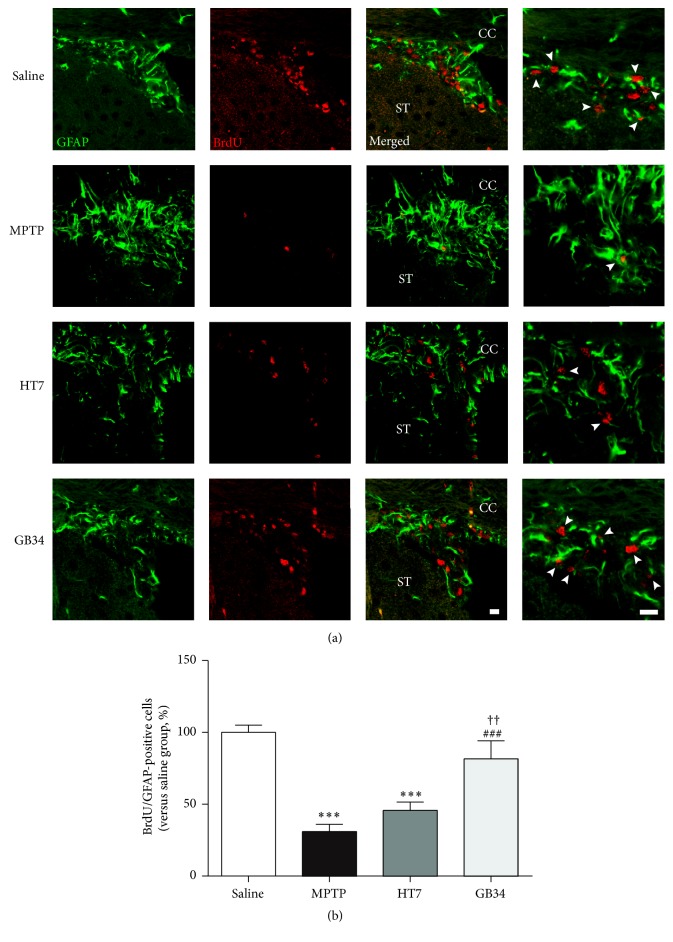
Figure 5.
Histological images and graphs of BrdU and glial fibrillary acidic protein- (GFAP-) positive cells in the SVZ. (a) BrdU and glial fibrillary acidic protein- (GFAP-) specific immunofluorescence staining. (b) The number of BrdU/GFAP-positive cells in the SVZ. Based on the results of BrdU (red) and GFAP (green)-positive cells in the SVZ, the number of the BrdU/GFAP-double stained cells in the SVZ decreased after MPTP administration, but acupuncture stimulation suppressed this decreased. CC: cerebral cortex. ST: striatum. Scale bar = 10 µm. Data are shown as the mean ± standard error of the mean. ∗∗∗P < 0.001 versus saline group, ###P < 0.001 versus MPTP group, and ††P < 0.01 versus HT7 group.

