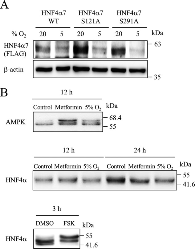
Figure 7.

Phosphorylation of HNF4α protein by hypoxia. A, MIN6 cells expressing wild-type (WT) or mutant (S121A or S291A) HNF4α7-FLAG by retrovirus transfection were cultured at either 5% O2 or 20% O2 for 24 h. Both exogenous and endogenous HNF4α proteins were examined using an anti-FLAG antibody and anti-HNF4α antibody, respectively. B, MIN6 cells were treated with metformin (2 mm) or incubated in 5% O2 for 12 and 24 h. Phosphorylation status of both AMPK and HNF4α was evaluated using Phos-tag SDS-PAGE. For a positive control, MIN6 cells were treated with either DMSO or forskolin (FSK, 100 μm) for 3 h.
