Figure 10.

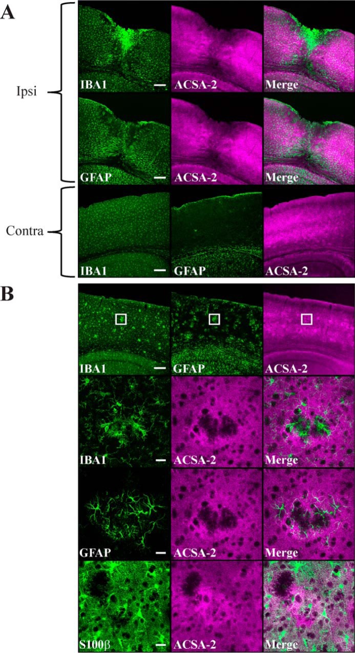
ACSA-2 staining is retained in reactive astrocytes. A, IBA1 (microglia), GFAP (reactive astrocytes), and ACSA-2 staining 5 days after a stab wound injury to the cortex. A low-magnification confocal image shows the pronounced migration of microglia ipsilateral (ipsi) to the injury. Astrocytes in the immediate vicinity of the injury appear to be lost, although those that remain appear to be highly reactive (as judged by up-regulation of GFAP expression throughout the ipsilateral cortex in comparison with the contralateral (contra) side). Note that ACSA-2 staining is still widespread even on reactive astrocytes, including those immediately adjacent to the site of injury. Tissue sections from three animals were analyzed with the same results. Scale bars, 200 μm. B, staining for IBA1, GFAP, S100β (astrocytes), and ACSA-2 in the cortex of an AppNL-G-F knock-in model of Alzheimer's disease at 6 months of age. Animals at this age show clear amyloid plaques in the cortex, which cause typical accumulation of microglia and local reactive astrogliosis, as seen in the low-magnification confocal images (top). Note that the ACSA-2 signal is consistent across the cortex. The boxed region is shown at higher magnification in the bottom panels. Plaques cause local aggregates of microglia to form in the tissue, which exclude astrocytes (as judged using an independent marker for astrocyte cell bodies, S100β). Astrocytes immediately adjacent to plaques show high levels of reactivity (as judged by up-regulation of GFAP expression) but do not lose immunoreactivity for ACSA-2. Tissue sections from two animals were analyzed with the same results. Scale bars, 200 μm (low magnification) and 20 μm (high magnification).
