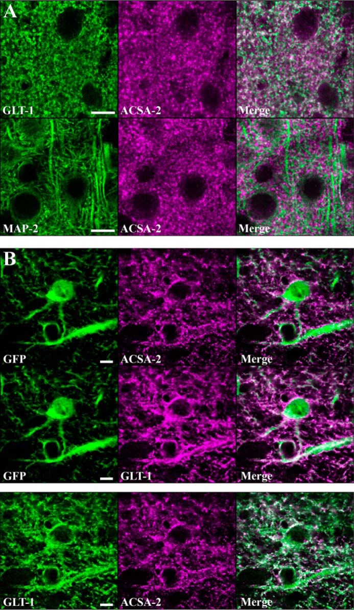
Figure 9.

ATP1B2 is expressed in discrete subcellular domains on the plasma membrane of astrocytes. A, top panels, astrocytes were stained with ACSA-2 and an antibody against an astrocyte-specific membrane protein, GLT-1. Imaging in the visual cortex revealed the staining patterns to be highly similar although non-overlapping. Bottom panels, co-staining of ACSA-2 together with a marker of neuronal microtubules (MAP2) showed highly dissimilar staining. Scale bar, 10 μm. B, top panels, tissue sections from an Aldh1l1-EGFP mouse. Images were taken in the corpus callosum, where the lower cell density allows the clear identification of individual astrocytes based on GFP expression. Both ACSA-2 and GLT-1 signal clearly localize to astrocytes. Note that ACSA-2 signal is detected around astrocyte cell bodies and extending into astrocyte processes. Scale bar, 5 μm. Bottom panels, both GLT-1 and ACSA-2 signal colocalize to the same cell, albeit in non-overlapping domains. Scale bar, 5 μm. Tissue sections from two animals were analyzed with the same results for both sets of stainings.
