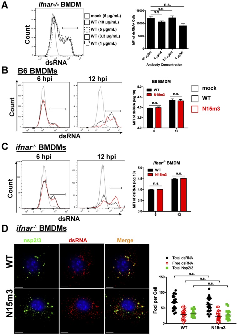
Fig. S6.
Evaluation of dsRNA levels in infected-macrophages. (A) ifnar−/− BMDMs were infected with WT virus at an MOI of 0.1. At 6 hpi, cells were stained with anti-dsRNA at stated concentrations and analyzed by flow cytometry. (B) B6 or (C) ifnar−/− BMDMs were infected with WT or N15m3 virus at an MOI of 0.1. At 6 and 12 hpi, cells were stained for dsRNA and analyzed by flow cytometry. BMDMs were sorted by removing debris in FSC-A/SSC-A plot, selecting single cells by FSC-H/FSC-A plot, and gating on dsRNA+ cells. (D) ifnar−/− BMDMs were infected with WT or N15m3 at MOI of 0.1, fixed at 12 hpi, and stained with anti-dsRNA, anti-nsp2/3, and Hoescht 33342. The foci from 25 images were counted using IMARIS software program. Values were analyzed by a unpaired t test and n.s. stands for not significant. Data are representative of two to three independent experiments. (Scale bars, 5 μM.)

