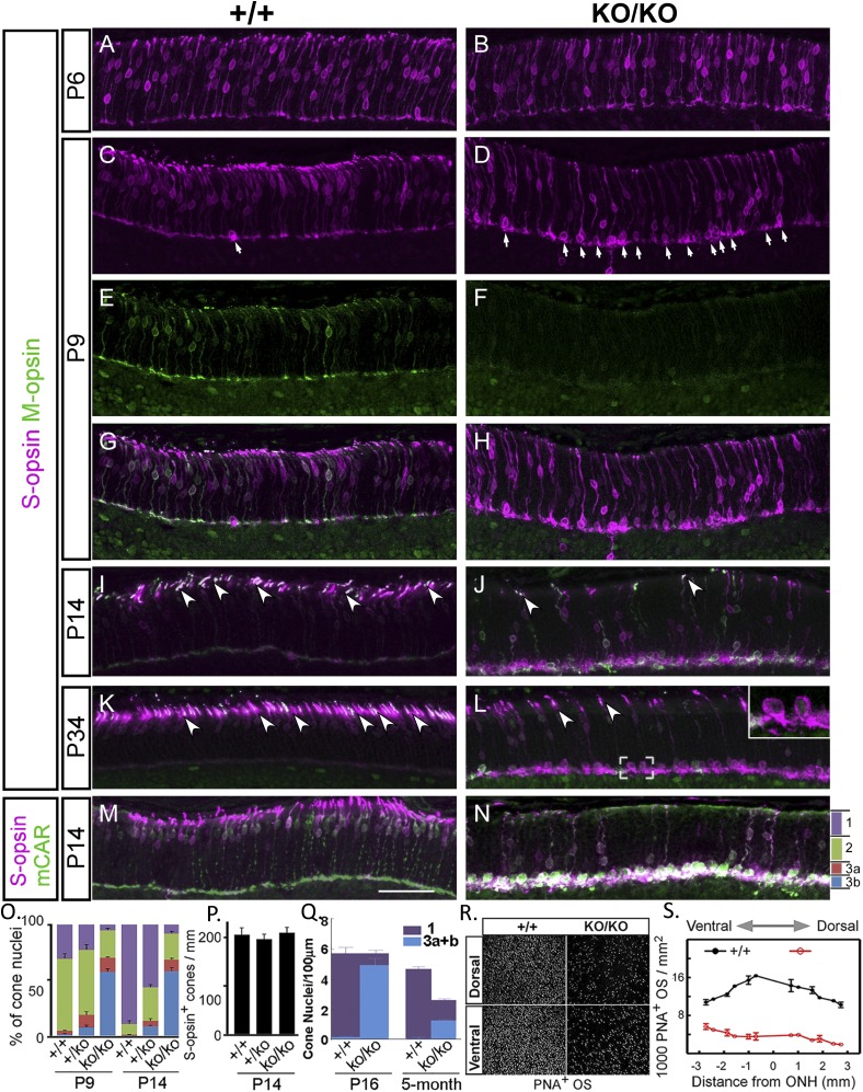
Fig. 3.
Aberrant nuclear positioning and outer segment formation in cone photoreceptors. (A–N) Representative images of immunostaining for S-opsin, M-opsin, and cone arrestin (mCAR) in retina (inferior) at postnatal ages indicated. ONL is divided into four bins: 1 apical third (purple), 2 middle third (green), with the basal third equally divided into 3a (red) and 3b (blue) (N). (O) Distributions of S-opsin+ nuclei in the four bins at P9 and P14, respectively (percentage). (P) Number of cone nuclei (S-opsin+) per millimeter of ONL length at P14 (P > 0.05). (Q) Number of cone nuclei per micrometer of ONL length as determined by the unique cone nuclear morphology on thin plastic sections (0.5 µm) at 5 mo (P < 0.05) under high magnification. Bins are similarly defined as in N, except that blue indicates the bottom third combining the two basal subdivisions (bins 3a+b). (R) Representative images of PNA staining of flat mounts of dorsal and ventral retina at P14. (S) Distributions of PNA staining throughout retina at P14 over distances from optical nerve head (ONH). (Scale bar, 50 µm in all panels.) Error bar is SEM.

