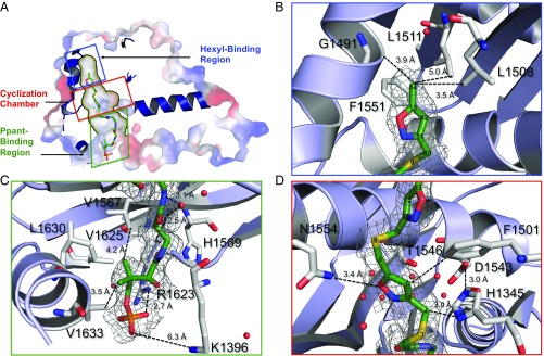
Fig. 3.
Structure of the 6c • PksA PT complex. (A) Surface rendition depicting the hexyl-binding chamber, crystallization chamber, and Ppant binding region within the 1.8 Å crystal structure of the 6c • PksA PT complex. The PPant-tethered heptaketide outlines various residues in the catalytic pocket. Schematic representation illustrates the close contacts in the (B) hexyl-binding region, (C) PPant binding region, and (D) cyclization chamber. Residue numbers and distances between the substrate and protein are provided.

