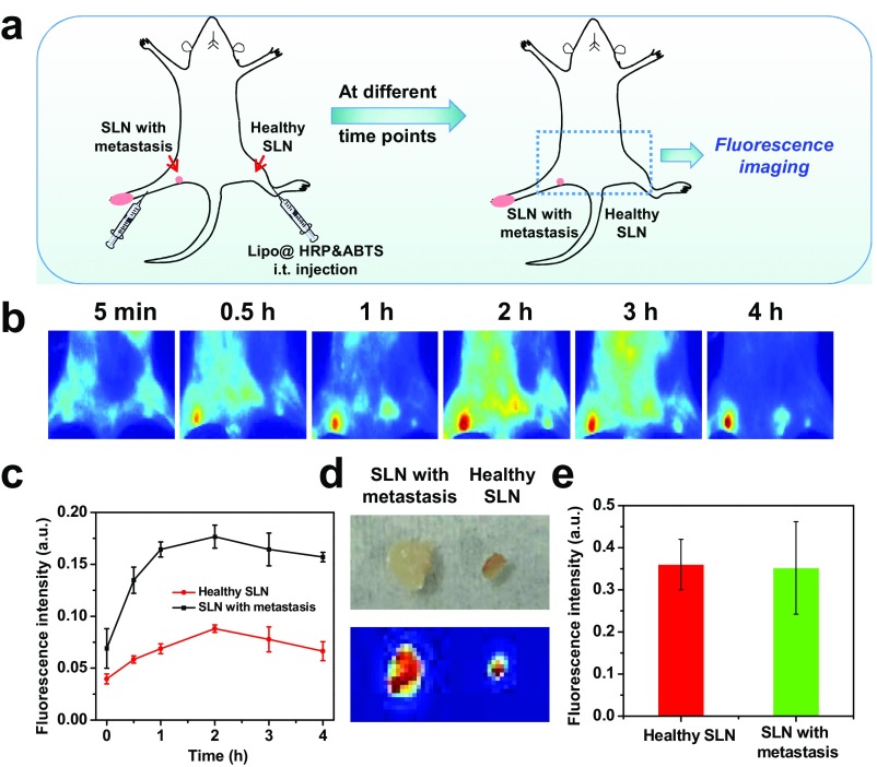
Fig. S8.
In vivo fluorescence imaging for SLN mapping. (A) Schematic illustration showing in vivo fluorescence imaging of SLNs of mice after local injection of Lipo@HRP&ABTS labeled with DiR, a lipophilic NIR fluorescent dye. (B and C) In vivo fluorescence images (B) and SLN fluorescence intensities (C) taken at different time points local injection of DiR-labeled Lipo@HRP&ABTS into mouse legs. (D) Ex vivo bright field (Top) and fluorescence (Bottom) images of metastatic lymph nodes and nonmetastatic nodes (on the opposite side) after local injection of DiR-labeled Lipo@HRP&ABTS into legs at both sides. (E) Relative fluorescence intensities of the SLNs based on fluorescence images shown in D. Although nonmetastatic lymph nodes with smaller sizes buried under a deeper location showed weaker in vivo fluorescence compared with metastatic nodes with larger sizes (due to a light penetration issue), the retention of liposomal nanoprobes in both types of lymph nodes seemed to be similar based on ex vivo imaging data.

