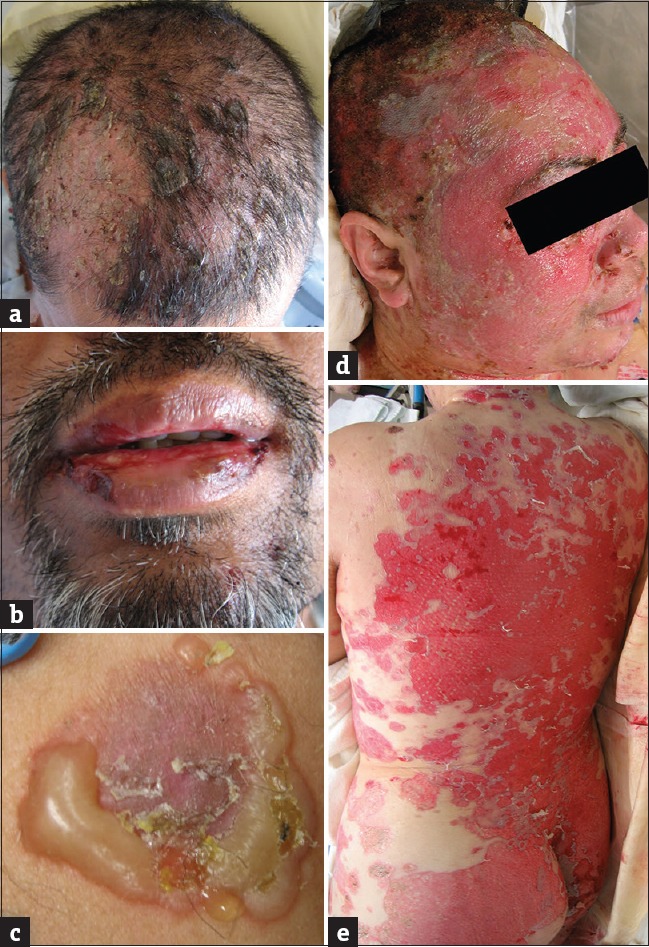
Figure 1.

Clinical photos of pemphigus lesions. (a) Flaccid bullae involving the scalp. (b) Erosions of oral cavity following blister rupture. (c) Characteristic flaccid blister with previously ruptured areas. (d and e) Widespread cutaneous erosions following rupture of bullae
