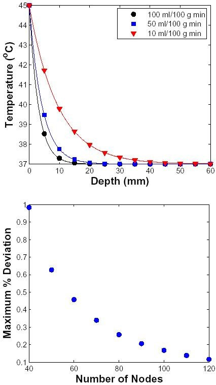
Figure 2.

Validation using one-dimensional geometry. The validation model is a homogeneous section of a material (here dermis) with uniform perfusion (homogeneous sink), 10 cm in length and 100 μm × 100 μm in area. The surface of the tissue was elevated to 45°C at t = 0 s. The perfusion level (in ml/100 g min) was varied as shown in inset. The solid line represents the analytical solution (Eq. 8) and the symbols represent the transport lattice solution. Top: Steady-state temperature as a function of depth. The 10 cm long tissue is represented by 100 lattice elements, but the temperature profile is shown only to the depth of 6 cm. Bottom: The 10 cm long tissue is now represented by different number of lattice elements. Maximum % deviation as a function of the number of nodes used to represent the tissue is shown. The deviation is computed as the maximum discrepancy between the simulated temperature and the corresponding analytical value normalized to the step increase in temperature (here = 8°C).
