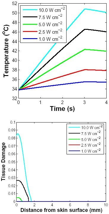
Figure 5.

Effect of applied power level for spatially distributed heating. Response to a 10 GHz microwave pulse of 3 s duration with four different power densities (inset in W cm-2). The layer of air farthest from the skin (2 mm) was at 25°C, the skin surface was at 34°C before the pulse was applied and the core temperature was fixed at 37°C. The blood perfusion level was assumed to be 10 ml/100 g min. Top: Surface temperature as a function of time. Bottom: Tissue damage indicator, Ω, as a function of depth. Only the two highest levels of power generate noteworthy values of Ω (the plots for lower power levels are, therefore, not visible in the figure).
