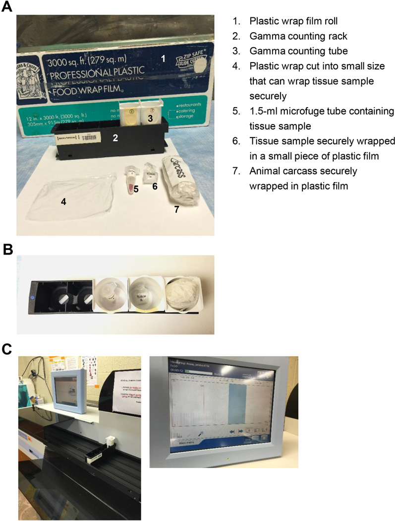
Figure 2. Illustration of radioactive tissue handling and loading to gamma counting tubes prior to measurement by a gamma counter.
A. Materials required for tissue sample wrapping. B. Top view of the gamma counting rack with previously wrapped samples loaded in each gamma counting tube. C. The gamma counting rack containing the sample tube is placed on the rack belt of the gamma counter, ready to be loaded inside the gamma detector (left panel). The gamma counter screen showing real-time radioactivity being detected from the tissue sample (right panel).

