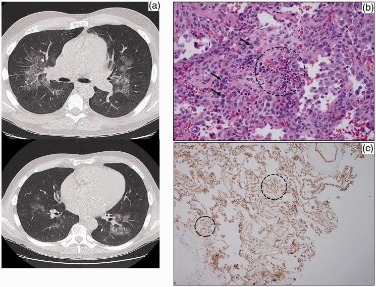
Fig. 2.
(a) Axial CT image shows bilateral GGOs with underlying interlobular septal thickening – the “crazy paving” sign. (b) Erythroid colony (circle) with myeloid precursors (arrows point out representatives) in the wall of airway. (c) CD31 immunostain showing increased small vessel density (circles).

