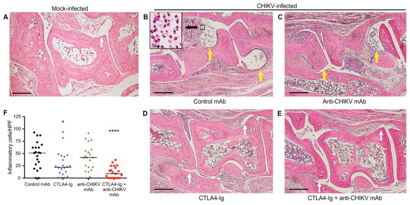
Fig. 5. Representative H&E staining of the ipsilateral midfoot joints in CHIKV-infected mice treated with CTLA4-Ig and anti-CHIKV mAb.
(A to F) Mice were inoculated with either PBS (A; mock) or 103 FFU of CHIKV (B to F) via a subcutaneous route. Animals were sacrificed, and histology of the ipsilateral foot was performed on day 7 after infection. CHIKV-infected mice received at day 3 a single intraperitoneal injection of 600 μg of isotype control antibody (B), 300 μg of anti-CHIKV mAb (C), 300 μg of CTLA4-Ig (D), or a combination of 300 μg of CTLA4-Ig and 300 μg of anti-CHIKV mAb (E). The number of inflammatory cells per HPF in the midfoot synovial space was quantitated in a blinded fashion (F). Results are representative of at least two independent experiments with n = 4 per treatment group and two sections assessed per foot. Scale bars, 200 μm. Yellow arrows, moderate to severe synovitis; white arrows, absent or mild synovitis *P < 0.05, ****P < 0.0001 (Kruskal-Wallis with Dunn’s post hoc analysis).

