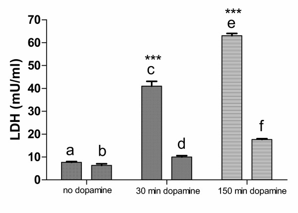
Figure 4.

Dopamine-induced cellular damage. Cells were incubated for 150 min with and without 100 nM dopamine and LDH-release was determined. (a) untreated control; (b) untreated cells kept for 30 min in normoxia followed by 120 min in xenon-atmosphere; (c) cells under normoxic conditions treated for 30 min with 100 nM dopamine followed by 120 min in normal medium; (d) cells under normoxic conditions treated for 30 min with 100 nM dopamine, followed by 120 min incubation in xenon-atmosphere in normal medium without dopamine; (e) cells under normoxic conditions treated for 150 min with 100 nM dopamine; (f) cells under normoxic conditions treated for 30 min with 100 nM dopamine, followed by 120 min incubation in xenon-atmosphere in medium containing 100 nM dopamine. No significant difference was found between (a) and (b) whereas the difference between (c) and (d), and (e) and (f) was highly significant (P < 0.0001).
