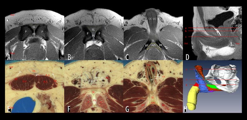
Figure 1.
Cross-sectional images of the suspensory ligamentous system and its adjacent structures on the VHM and MRI images. (D) Positions of the transverse sections of panel A–C. Sections show the medium-signal-intensity suspensory ligament (A, B) and arcuate pubic ligament (C) on the MRI images. (H) Positions of the transverse sections of panel E–G. Sections show the position of the fundiform ligament (A, B), suspensory ligament (B), and arcuate pubic ligament (C) in VHM data set. SF – Scarpa’s fascia; FL – fundiform ligament; LA – linea alba; RA – rectus abdominis; SL – suspensory ligament; PS – pubic symphysis; P – pubis; DF – deep fasica of the penis; CC – corpora cavernosa; TA – tunica albuginea; APL – arcuate pubic ligament; RP – ramus of pubis.

