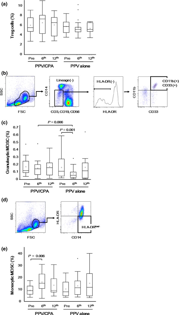
Figure 4.

Inhibitory immune cells before and after personalized peptide vaccination (PPV). Inhibitory immune cells, including Treg cells, granulocytic myeloid‐derived suppressor cells (MDSC), and monocytic MDSC, in peripheral blood mononuclear cells (PBMC) were examined before and after PPV (6th and 12th vaccination). (a) The percentages of Foxp3+ CD25+ cells in CD4+ cells were determined before and after PPV. (b) In the cell subset negative for lineage markers (CD3, CD19, CD56, CD14) and HLA‐DR in the lymphocyte/monocyte gate, granulocytic MDSC were identified as positive for CD33 and CD11b. (c) The percentages of CD11b+ CD33+ cells in PBMC were determined before and after PPV. (d) Monocytic MDSC were identified as positive for CD14 and low (or negative) for HLA‐DR in the monocyte gate. (e) The percentages of HLA‐DR low/− cells in CD14+ cells were determined before and after PPV. Box plots show median and interquartile range (IQR). The whiskers (vertical bars) are the lowest value within 1.5× IQR of the lower quartile and the highest value within 1.5× IQR of the upper quartile. Data not included between the whiskers were plotted as an outlier with dots. “X” shows the mean of the data.
