
Figure 2.
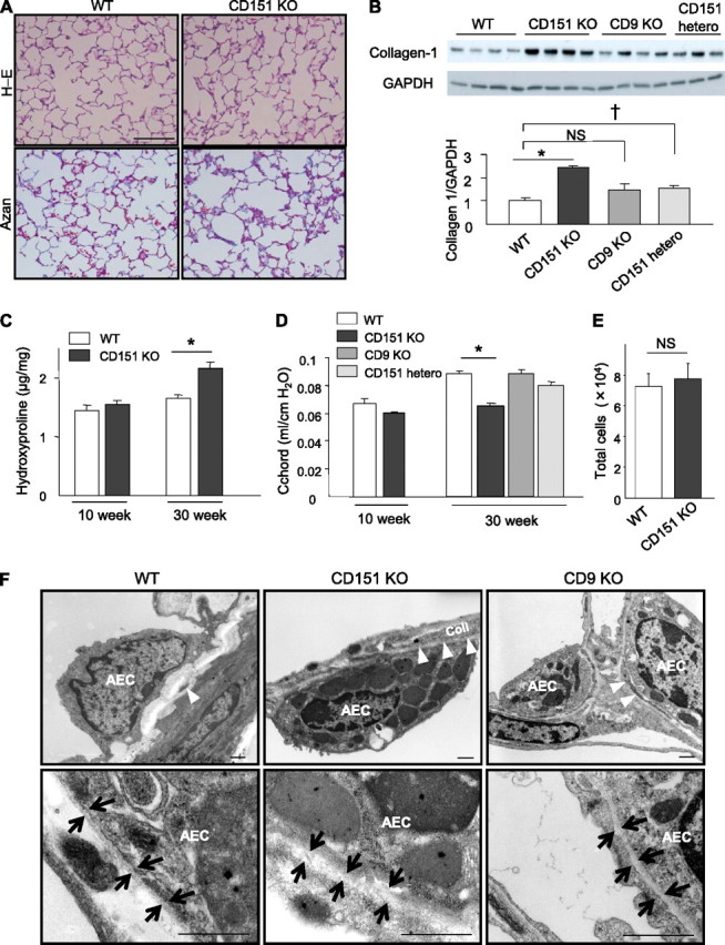
CD151 knockout (KO) mice spontaneously develop age-related pulmonary fibrosis. (A) Representative lung sections from wild-type (WT) and CD151 KO mice at 30 weeks of age stained with hematoxylin-eosin (H-E) and Azan. Bar = 100 μm. (B) Immunoblotting of total lung homogenate revealed increased expression of collagen-1 in CD151 KO and heterozygous (hetero) mice but not in CD9 KO mice compared with WT mice at 30 weeks of age. *P < 0.001 (WT vs. CD151 KO), †P < 0.05 (WT vs. CD151 hetero). (C) Hydroxyproline contents of total lungs were measured and normalized against lung weight (μg/mg, n = 5 per group). *P < 0.05. (D) Lung compliance was measured by a quasistatic pressure/volume maneuver, and is shown as C chord values (n = 4−5 per group). *P < 0.001. (E) Total cell number in bronchoalveolar lavage fluids was comparable between CD151 KO and WT mice at 30 weeks of age (n = 4 per group). (F) Electron microscopy of CD151 KO lung at 16 weeks of age revealed increased collagen (coll) deposition in alveolar walls and hypertrophied type II alveolar epithelial cells (AECs) with many lamellar body−like structures. Basement membranes (BM) under these AECs were irregularly thickened. The number of lamellar body−like structures was also increased in CD9 KO lungs but not as prominently as in CD151 KO lungs. Arrowheads indicate coll in alveolar walls; arrows indicate BM. Bar = 1 μm.