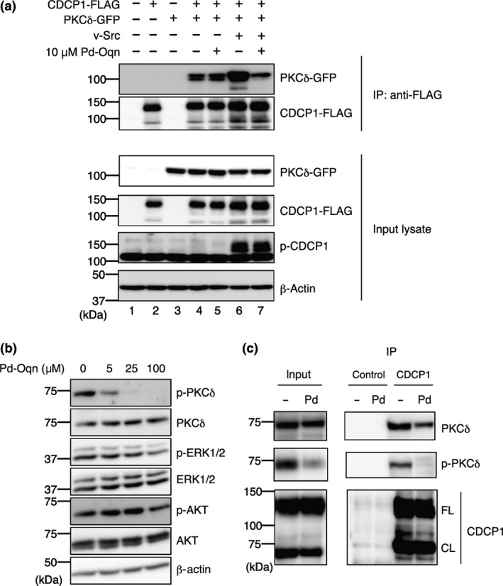
Figure 3.

Inhibition of the CDCP1‐PKCδ pathway by Pd‐Oqn. (a) Blockage of the CDCP1‐PKCδ interaction by Pd‐Oqn in cells. Immunoprecipitation was performed as described in Materials and Methods. Immunoprecipitated proteins and input lysates were subjected to western blotting with the antibodies as indicated at the right side. The top indicates the transfection plasmids and Pd‐Oqn addition. (b) Inhibition of the phosphorylation of PKCδ by Pd‐Oqn. 44As3 cells were treated with Pd‐Oqn and the lysates were subjected to western blotting with the antibodies as indicated at the right side. (c) Inhibition of the endogenous CDCP1‐PKCδ interaction by Pd‐O qn. 44As3 cells were treated with DMSO (‐) or Pd‐Oqn (Pd) for 24 h and the cell lysates were subjected to immunoprecipitation with anti‐CDCP1 antibody or control IgG. The precipitated proteins (IP) and the cell lysate (input) were subjected to western blotting with the antibodies as indicated at right side. CDCP1 bands showed both full‐length (FL) and cleaved form (CL) at about 135 and 70 kDa respectively. Molecular weight is indicated at the left.
