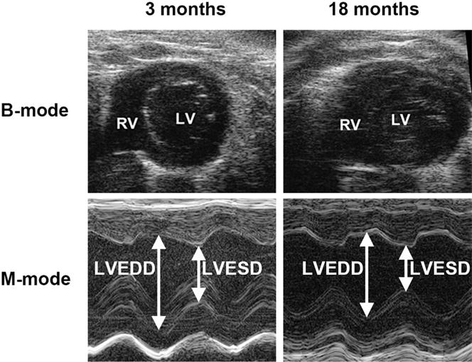
Fig. 4. Representative M-mode echocardiograph images obtained from mice at ages of 3 months and 18 months.

RV, right ventricle; LV, left ventricle; LVEDD, left ventricular end diastolic dimension; LVESD, left ventricular end systolic dimension.

RV, right ventricle; LV, left ventricle; LVEDD, left ventricular end diastolic dimension; LVESD, left ventricular end systolic dimension.