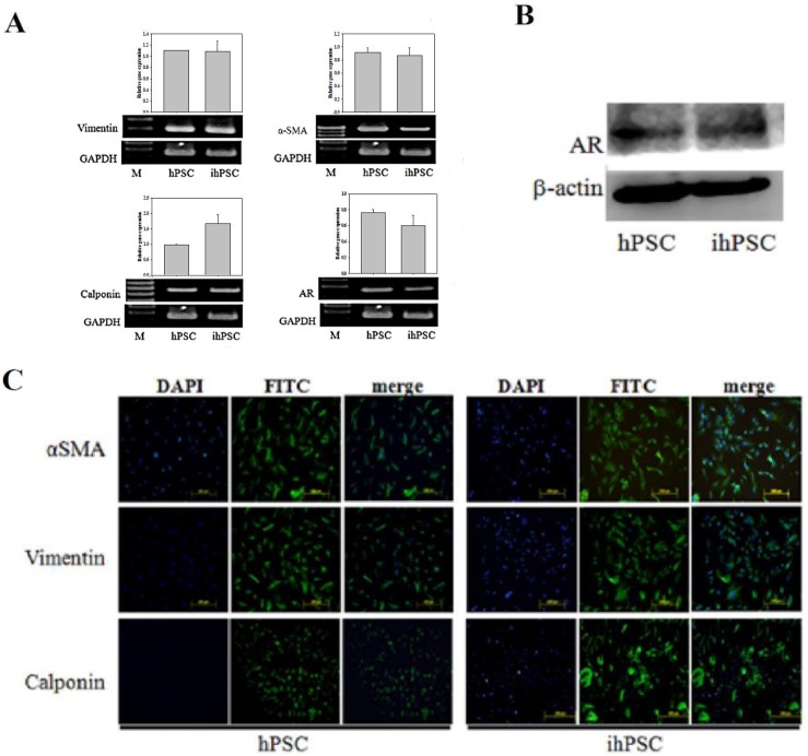
Fig 3. Phenotypic characteristics of ihPSC.
(A) RT-PCR analysis of specific mRNA transcripts of cell markers, including vimentin, α-smooth muscle actin (α-SMA), calponin, and androgen receptor (AR) and their relative quantification. GADPH was used as internal control. (B) Western blot analysis for proteins isolated from hPSC and ihPSC cells confirmed the expression of AR with anti-AR antibody. β-actin was used as the internal control. (C) Immunocytofluorescence staining for α-SMA, vimentin, and calponin (using FITC labeled antibodies). Nuclei were counterstained with DAPI (blue). FITC and DAPI stainings were merged to show the localization of specific proteins. Scale bar, 200 μm. No significant difference was observed among cell markers during RT-PCR analysis.

