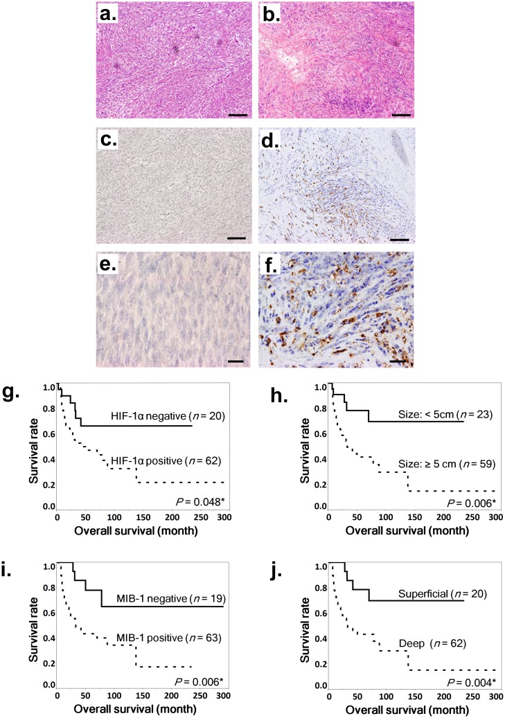
Fig 1. Hematoxylin and eosin staining and immunohistochemical staining of HIF-1α in MPNST samples and association between nuclear HIF-1α expression and poor prognosis in MPNSTs.
a–f. Staining with hematoxylin and eosin (a and b) and immunohistochemical staining of HIF-1α (c–f) in MPNST specimens. Representative cases: a HIF-1α–negative specimen (a, c, and e) and a HIF-1α–positive specimen (b, d, and f). Scale bar, 100 μm in a–d and 20 μm in e and f. g–j. Kaplan-Meier survival curves for all patients based on positive or negative nuclear HIF-1α expression (g), tumor diameter of 5 cm or more (h), positive or negative MIB-1 expression (i), and deep tumor location (j). Log-rank tests were performed to determine statistical significance.

