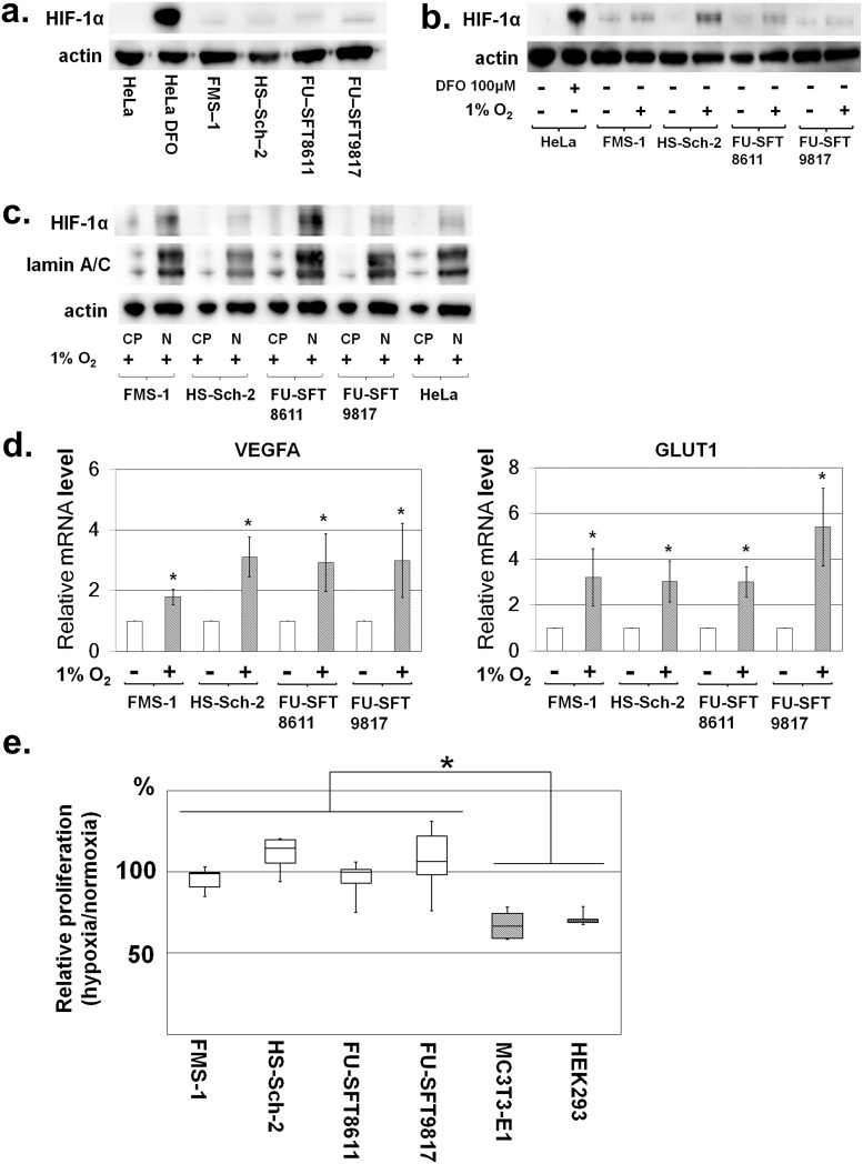
Fig 2. Nuclear expression of HIF-1α and MPNST cell line proliferation under normoxic and hypoxic conditions.
a. Nuclear expression of HIF-1α in MPNST cell lines under normoxia was confirmed by Western blotting of HIF-1α using nuclear extract from each cell line. In HeLa (cervical cancer cell line) cells, the expression of HIF-1α is shown under hypoxia caused by the addition of DFO. Meanwhile, definite expression of HIF-1α was observed in all MPNST cell lines. b. Nuclear expression of HIF-1α in MPNST cell lines was induced by hypoxia. Actin was used for internal normalization. c. Based on comparison with nuclear protein lamin A/C, HIF-1α was mainly localized in the nuclei. In Fig 2c, “CP” represents cytoplasmic proteins, and “N” represents nuclear proteins. d. VEGFA and GLUT1 expression downstream of HIF-1α was enhanced under hypoxia. e. Cell proliferation under normoxic and hypoxic conditions in MPNST cell lines. Growth at 48 h after cell seeding was compared under normoxia and hypoxia. In the hypoxic condition, cell proliferation of non-transformed cell lines (MC3T3-E1 and HEK293) was suppressed; however, growth of MPNST cell lines under hypoxia was comparable to that under normoxia. Experiments were performed six times. Data in graphs are presented as box-and-whisker plots, and statistical comparisons were performed using the Mann-Whitney U test. *P <0.05.

