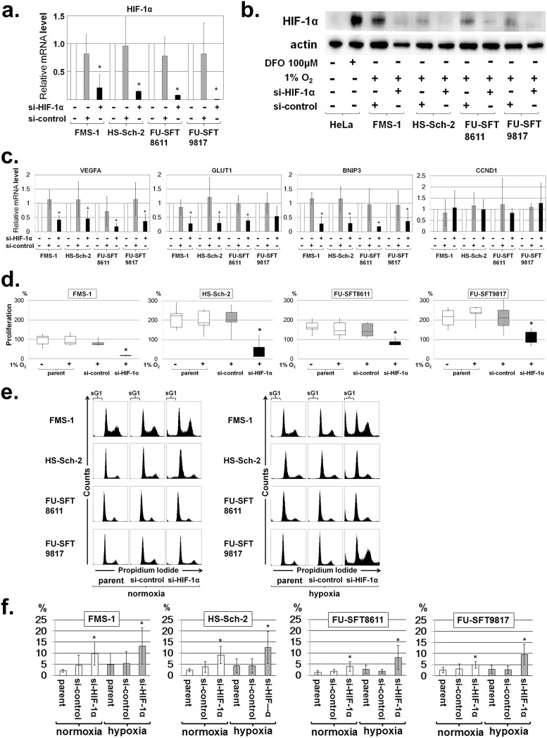
Fig 3. Knockdown of HIF-1α by si-RNA and effects of si-HIF-1α on cell proliferation and cell cycle progression.
Assessment of HIF-1α knockdown in MPNST cell lines by the following: a. Real-time quantitative PCR (mean ± SD; *P < 0.05) under normoxia; and b. Western blotting of HIF-1α after induction of HIF-1α–specific si-RNA under hypoxia. The si-HIF-1α suppressed the expression of HIF-1α compared to control si-RNA. Actin was used for internal normalization. c. Downregulation of downstream genes by si-HIF-1α. VEGFA, GLUT1, and BNIP3 expression downstream of HIF-1α was decreased by si-HIF-1α, whereas expression of CCND1 downstream of HIF-2α remained unchanged. d. Effects of si-HIF-1α on cell proliferation in MPNST cell lines. Knockdown of HIF-1α by si-RNA suppressed the proliferation of MPNST cell lines under hypoxia. Data in graphs are presented as box-and-whisker plots, and statistical comparisons were performed using the Mann-Whitney U test. *P < 0.05. e and f. Effects of si-HIF-1α on cell cycle progression in MPNST cell lines. e. Representative cell cycle profile of MPNST cell lines after knockdown of HIF-1α by si-RNA. The areas labelled “sG1” in the figures represent subG1 fractions. f. SubG1 fractions increased by HIF-1α knockdown in MPNST cell lines under normoxia and hypoxia. Experiments were performed in triplicate or more, and data are expressed as the mean ± SD. *P < 0.05. Each value is listed in S1 Table.

