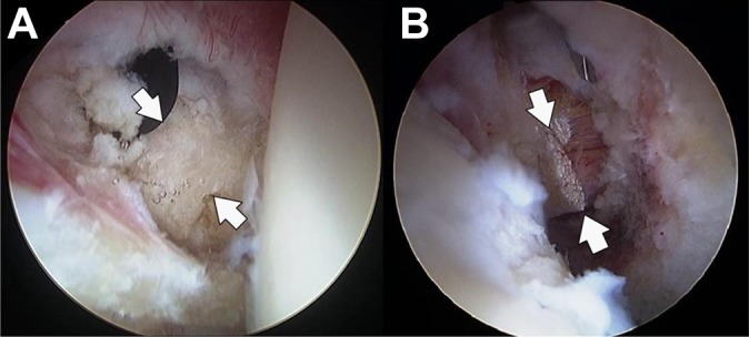
Figure 1.

Arthroscopic views of the right hip showing the exposure and release of the iliopsoas tendon at the level of the labrum. (A) Prior to the release, an anterior capsulotomy was performed to expose and define (arrows indicate the edges of the tendon) the borders of the tendon. (B) The tendon was then released with a combination of the radiofrequency device and a beaver blade. Care was taken to only release the tendinous portion of the iliopsoas muscle-tendon unit, which, based on a prior study, leaves 60% of the muscle-tendon unit (the muscular portion) intact.2,6
