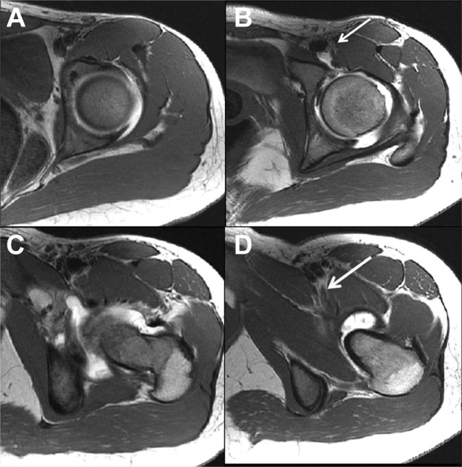
Figure 2.

Pre- and postoperative axial T1-weighted hip magnetic resonance arthrography (MRA) images of a 16-year-old female with left hip pain. (A and C) Preoperative images demonstrated no muscle atrophy. (B and D) Postoperative images demonstrated grade 4 atrophy of the psoas muscle above the level of the acetabular rim (arrow, B) and grade 1 atrophy below the level of the acetabular rim (arrow, D). The postoperative MRA was performed 13 months after the patient’s labral-level iliopsoas tenotomy, and her modified Harris Hip Score at that time was 82.5 points.
