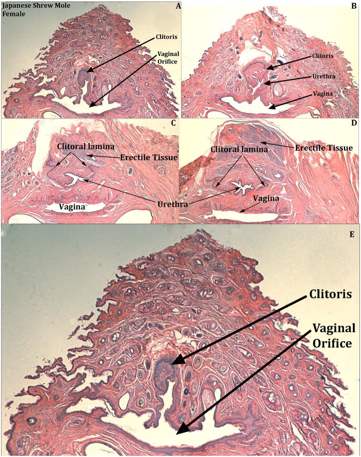
Figure 15.
Transverse H&E stained sections of adult Japanese shrew mole (Urotrichus talpoides) female genitalia from the breeding season identifying the distal beginning of the clitoris and the connection of the urethra to the vaginal vestibule (E). Sections (A–D) progress distal to proximal from left to right. In (A & E) note that the U-shaped clitoral lamina is fused to the urethra, which opens into the vaginal orifice. In (B) the urethra is separate from the vagina. In (C & D) the U-shaped clitoral lamina is represented merely as its right and left pieces that flank the urethra and dorsal to the urethra is diffuse erectile tissue.

