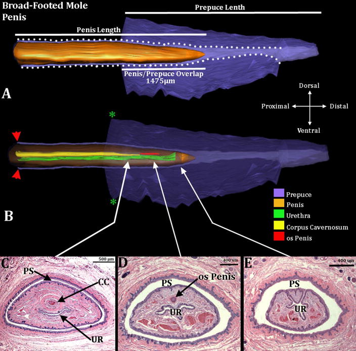
Figure 2.
Three-dimensional reconstruction of the prepuce and penis of an adult broad-footed mole (Scapanus latimanus) with transverse sections depicting morphology at different locations within the penis. Three-dimensional reconstruction (A) the prepuce is semi-transparent with the penis opaque orange demonstrating the shape and the position of penis relative to the prepuce. The mole penis at rest is an “internal” organ housed within the preputial space (lighter purple region bordered by white dotted lines). Three white lines are labeled denoting the length of the external prepuce, the length of the penis and the length the penis/prepuce overlap. Three dimensional reconstruction (B) the prepuce and penis are rendered semi-transparent to show the internal morphology of the penis. The perineal appendage (prepuce) merges with the body surface (green asterisks). Red arrowheads denote the reflection of the inner preputial epithelium onto the surface of the penis. The distal end of the urethra (green) is the urethral meatus where urine/semen exits from the penis. Three transverse section images (C–E) depict the internal morphology of the penis from proximal to distal at differing morphological points. CC=Corpus Cavernosum, PS=Preputial Space, UR= Urethra. Note the morphometric measures mentioned in the text.

