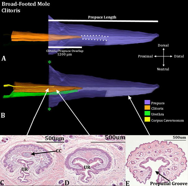
Figure 3.
Three-dimensional reconstruction of the prepuce and clitoris of an adult broad-footed mole (Scapanus latimanus) with transverse sections depicting morphology at different locations within the clitoris. Three-dimensional reconstruction (A) the prepuce is semi-transparent with the clitoris opaque orange demonstrating the shape and the position of clitoris relative to the prepuce. The mole clitoris is housed within the prepuce (in purple); lighter purple depicts the preputial grove distally and when bordered by white dotted lines depicts the preputial space. The 2 white lines denote the prepuce length and the length of the clitoris/prepuce overlap. Three-dimensional reconstruction (B) the prepuce and clitoris are rendered semi-transparent to show the internal morphology of the clitoris. The perineal appendage (prepuce) merges with the body surface (green asterisks). Three transverse H&E section images (C–E) depict the morphology of the clitoris and prepuce at differing morphological points from proximal to distal. CC=Corpus Cavernosum, UR=Urethra. Note the morphometric measures mentioned in the text.

