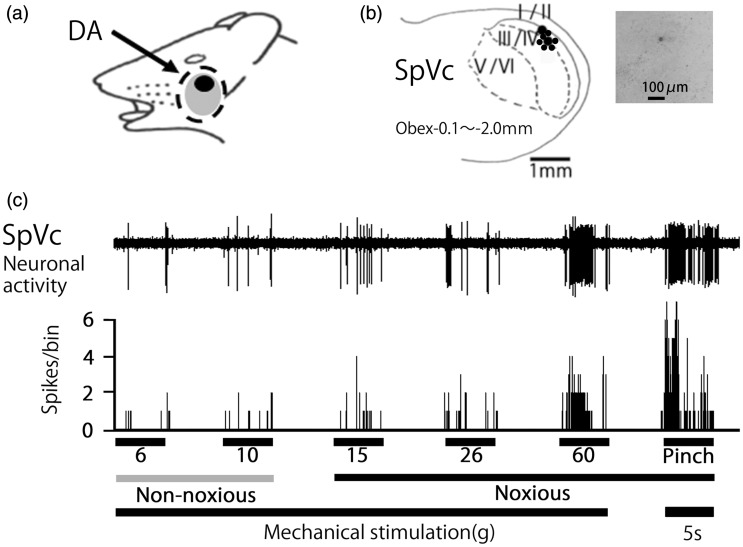
Figure 2.
General characteristics of spinal trigeminal nucleus caudalis (SpVc) wide-dynamic range (WDR) neuronal activity in response to mechanical stimulation of orofacial skin. (a) Typical example of receptive field of whisker pad in the facial skin. Shaded area indicates the region applied with decanoic acid (DA). (b) Distribution of SpVc WDR neurons responding to non-noxious and noxious mechanical stimulation of the facial skin (n = 18). Inset: example for histological confirmation of recording site. The number below each drawing indicates the frontal plane in relation to obex. (c) Example of non-noxious and noxious mechanical stimulation-induced firing of SpVc WDR neurons.

