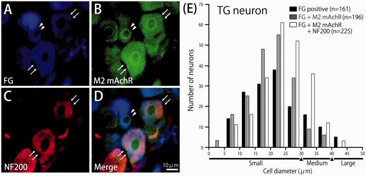
Figure 6.
Immunoreactivity of muscarinic acetylcholine M2 receptor (M2 mAchR) and neurofilament protein-200 (NF-200) in the trigeminal ganglion (TG) neurons innervating facial skin. (a) Floresence micrograph showing retrograde FG-labeled TG neurons innervating Facial skin. (b) M2 mAchR immunoreactive TG neurons in the same section. (c) NF-200 immunoreactive TG neurons in the same section. (d) Merged. A typical example of an FG-labeled TG neuron expressing M2 mAchR is indicated by filled triangles. A typical example of FG-labeled TG neurons co-expressing M2 mAchR and NF-200 are indicated by arrows. (e) Histogram showing the cell size spectrum of M2 mAchR and NF-200 positive FG-labeled TG neurons in the trigeminal ganglia.

