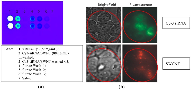
Figure 4.
(a) Cy-3-siRNA complexed to SWCNT is not fluorescent (lane 3); (b) H2122 NSCLC cells in culture exposed to Cy-3-siRNA/SWCNT 1 µg/mL for 6 h. Cells were washed and examined by bright field and fluorescent microscopy. Top views illustrate left: the H2122 cells in culture (10.6 pix/µm); right: intracellular Cy-3-siRNA by visible fluorescence (10.6 pix/µm); and bottom views left: illustrate the same H2122 cells in culture (2 pix/µm); right: the intracellular SWCNT by NIR fluorescence (2 pix/µm) in the same cells. All cells in culture contained both the siRNA and SWCNT. Control cells not exposed to Cy-3 siRNA/SWCNT complex showed no fluorescence under either condition (data not shown).

