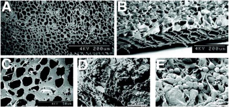
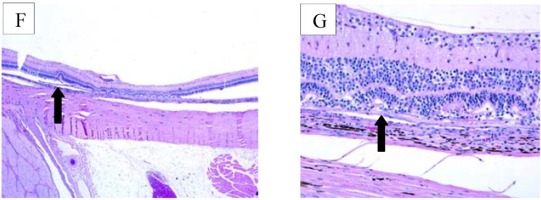
Figure 1.
PLGA-PLLA Co-polymer. Scanning Electron Micrographs, adapted from Tomita et al. 2005 [26], depict the PLLA-PLGA copolymer before (A–C) and after RPC seeding (D,E). Below are representative examples of histology from transplant eyes 30 days after transplantation. The arrow of the left image (F) depicts the location of the polymer in the subretinal space. The arrow on the right (G) shows a disruption of the retinal layers due to an immune-like response.


