
Figure 4.
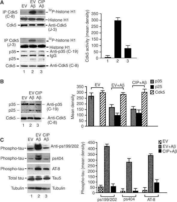
CIP inhibited Aβ1−42-induced endogenous p25/Cdk5 hyperactivity and tau hyperphosphorylation in cortical neurons. (A) 7-DIC cortical neurons were infected with EV or CIP and treated with 10 μM Aβ1−42 for 6 h. Cdk5 was immunoprecipitated from equal amounts of lysates with polyclonal C-8 and J-3 antibodies, and subjected to histone H1 kinase assays. The bar graph represents average mean optical density±s.e.m. of phospho-histone levels in four separate experiments. Coomassie panels show equal amounts of histone present in the assay. To determine Cdk5, p25 and p35 levels in the Cdk5 immunoprecipitates, Cdk5 was immunodetected using polyclonal J-3 antibody in the C-8 IP. In the J-3 IP, p25 and p35 were immunodetected using polyclonal C-19 antibody; Cdk5 was detected using rabbit polyclonal C-8. (B) Western blots were performed to detect the presence of p35, p25 and Cdk5 using lysates from (A). The bar graph shows mean density±s.e.m. measurements of each of the three proteins of four independent experiments. (C) Lysates from (A) were resolved by SDS–PAGE. Lane 1: EV; lane 2: EV+Aβ1−42; lane 3: CIP+Aβ1−42. The top three panels show phospho-tau using different phospho-tau antibodies (anti-pS199/202, anti-pS404 and AT8), the fourth panel shows total tau (Tau5) and the bottom panel shows tubulin. The bottom two panels show equal amounts of total tau (Tau5) and tubulin in the lysates. The bar graph shows mean optical densities of phospho-tau ps199/202, ps404 and AT-8 of four separate experiments.