Figure 6.

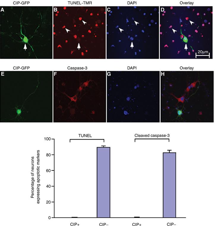
CIP reduces neuronal apoptosis induced by Aβ1−42 treatment in cortical neurons as measured by TUNEL assay and ICC assay of cleaved caspase-3. p35 Promoter-CIP-hrIRES-GFP(CIP-GFP) and p35 Promoter-empty vector hrIRES-GFP (GFP) were transfected into 3-DIC cortical neurons and after 24 h neurons were treated with 10 μM Aβ1−42 for 6 h. Neurons were fixed and stained in separate experiments for TUNEL and caspase-3 (cleaved) to show apoptotic neurons. In the first row (A–D), neurons were stained for TUNEL. (A) CIP-GFP; (B) TUNEL-TMR; (C) nuclear staining with DAPI; (D) overlay of CIP and TUNEL. In the second row (E–H), neurons are stained for cleaved caspase-3 expression. (E) CIP-GFP; (F) cleaved caspase-3; (G) nuclear staining with DAPI; (H) overlay of CIP and cleaved caspase-3. Cell counts were performed as follows: 10 independent fields were analyzed with a total of 500 neurons where TUNEL and cleaved caspase-3 could be counted. DAPI staining gave the total number of neurons and CIP was identified by GFP. The bar graph shows the quantity of neuronal apoptosis expressed as mean±s.e.m. from four separate transfections.
