
Figure 1.
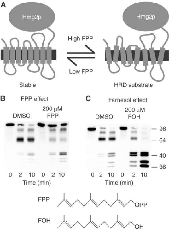
Farnesol but not FPP increased Hmg2p-GFP proteolysis rate. (A) Structural transition model of Hmg2p-regulated degradation. The large catalytic domain is on the cytosolic face of ER. (B) Effect of FPP. Microsomes bearing Hmg2p-GFP were resuspended in buffer with DMSO or 200 μM FPP, and then incubated with trypsin (15 μg/ml) at 30°C. Samples were removed at the indicated times and evaluated by 14% SDS–PAGE and immunoblotting of the lumenal Myc epitope. (C) Effect of farnesol (FOH). Microsomes bearing Hmg2p-GFP were resuspended in buffer with DMSO or 200 μM FOH, and then incubated with trypsin and evaluated as described above. Molecular weight markers (in kDa) are shown.