Figure 4.
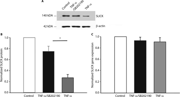
TNF-α decreases membrane expression of SLICK channels via p38 MAPK through possible posttranslational modification in DH neurons.
Notes: (A) Representative blot of membrane SLICK biotinylation assay. Membrane biotinylation and precipitation by streptavidin followed by Western analysis using a SLICK-specific antibody were performed on untreated neurons, neurons treated with 10 ng/mL TNF-α for 10 min, or neurons pretreated with 10 μM of p38 inhibitor SB202190 for 30 min followed by 10 ng/mL TNF-α for 10 min. (B) Densitometric analysis of SLICK membrane expression when normalized to β-actin. (C) Normalized SLICK gene expression levels. *P<0.05.
Abbreviations: DH, dorsal horn; TNF-α, tumor necrosis factor-alpha.

