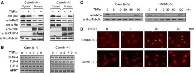
Figure 2.

Expression levels of components of the NF-κB signaling pathway and nuclear–cytoplasmic shuttling of NF-κB is not affected in Carm1(−/−) MEFs. (A) Protein expression levels of p65, c-Rel, RelB, p300 and PARP-1 are not affected in Carm1(−/−) MEF cells. Carm1(+/+) and Carm1(−/−) MEF cells were treated with TNFα (10 ng/ml) for 20 min and cytoplasmic and nuclear extracts resolved by SDS–PAGE followed by subsequent immunoblot analysis for p65, c-Rel, RelB, p300 and PARP-1 (A, left and right panels). α-Tubulin was used as an internal standard. (B) mRNA expression levels of TLR4, TLR2 and IRAK4 are not impaired in Carm1(−/−) MEF cells. Carm1(+/+) and Carm1(−/−) MEF cells were treated with TNFα (10 ng/ml) (B, left and right panels) and RNA isolated at the indicated time points followed by RT–PCR analysis for TLR4, TLR2, IRAK4 and HPRT (B, left and right panels). (C) Degradation and re-synthesis of IκBα is not affected in Carm1(−/−) MEF cells. Carm1(+/+) and Carm1(−/−) MEF cells were treated with TNFα (10 ng/ml) (C, left and right panels) and whole-cell extracts isolated at the indicated time points and resolved by SDS–PAGE, followed by subsequent immunoblot analysis for IκBα. α-Tubulin was used as an internal standard. (D) Nuclear translocation of NF-κB is not delayed in Carm1(−/−) MEFs. Carm1(+/+) and Carm1(−/−) MEF cells were treated with TNFα (10 ng/ml) (D, upper and lower panels) and fixed with paraformaldehyde at the indicated time points followed by immunostaining for p65 and analysis by immunofluorescent microscopy.
