FIGURE 3:
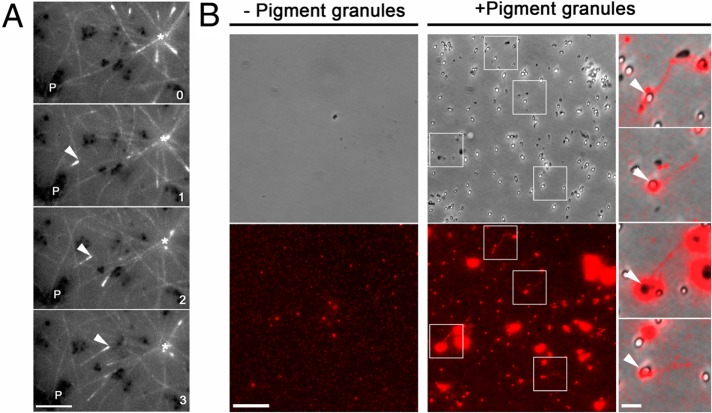
Pigment granules nucleate MTs in vivo and in vitro. (A) Sequential live fluorescence images of the centrosome area of an EGFP-EB1–expressing melanophore injected with TYRP1 antibodies and treated with melatonin; an asterisk indicates the position of the centrosome, P shows the location of a clump of pigment granules, and an arrowhead marks an EGFP-EB1 comet that emerges from the pigment granule clump and moves toward the centrosome; numbers indicate time in minutes. Birth of the MT tip labeled with EGFP-EB1 away from the centrosome and its growth toward the cell center suggest nucleation of the MT on a granule clump cross-linked with TYRP1 antibodies; bar, 2 μm. (B) Left and middle, phase contrast (top) and fluorescence (bottom) images of pellets of Cy3-labeled tubulin preparations incubated in the absence (left) or presence (right) of a suspension of purified pigment granules isolated from melanophores treated with melatonin to induce granule aggregation; right, overlays of boxed areas shown in the middle; tubulin pellets that were incubated in the presence of pigment granules contain short MTs that are often attached to pigment granules; bars, 10 μm (left and middle), 2 μm (right).

