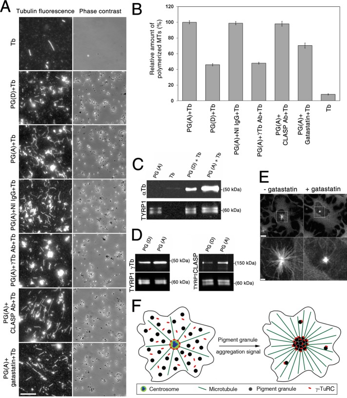
FIGURE 4:
Pigment granule aggregation signals increase nucleation of MTs on pigment granules, and this increase correlates with the recruitment of γ-tubulin. (A) Fluorescence (left) and phase contrast (right) images of pellets of immunostained MTs assembled by polymerization of twice-cycled tubulin incubated alone or with different pigment granule preparations; shown are images from (Tb) alone, (PG(D)+Tb) pigment granules isolated from cells treated with MSH to induce granule dispersion, (PG(A)+Tb) pigment granules from cells treated with melatonin to trigger aggregation and preincubated with buffer only, (PG(A)+IgG+Tb) pretreated with nonimmune rabbit IgG, (PG(A)+ γ-Tb antibody+Tb) pretreated with γ-tubulin antibodies, (PG(A)+CLASP antibody+ Tb) pretreated with CLASP antibodies, or (PG(A)+gatastatin+Tb) pretreated with γ-tubulin inhibitor gatastatin. Pigment granules increased the amount of assembled MTs, and this effect was greater in the case of granules isolated from melatonin- compared with MSH-treated cells; preincubation of granules with γ-tubulin antibodies or gatastatin but not with nonimmune IgG or CLASP antibodies reduced the amount of assembled MTs; bar, 10 μm. (B) Quantification of the number of assembled MTs in samples shown in A; each bar represents the average value of 30 measurements in two independent experiments; the data are expressed as percentage of average number of MTs assembled in the presence of pigment granules isolated from melatonin-treated cells and incubated with relevant buffer, which is taken as 100%; error bars are mean ± SEM. (C) Comparison of MT nucleation activity of pigment granules isolated from melanophores treated with MSH or melatonin by immunoblotting of granule pellets with α-tubulin antibodies; blots with α-tubulin (top) or TYRP1 (bottom; loading control) antibodies of pellets from samples containing pigment granules isolated from melatonin-treated cells (PG(A)), purified tubulin (Tb), or mixtures of purified tubulin with pigment granules isolated from MSH- or melatonin-treated melanophores (PG(D)+Tb, and PG(A)+Tb, respectively); α-tubulin bands are absent from pellets of pigment granules or tubulin preparations (an indication that pigment granule preparations do not contain significant amounts of tubulin and that MTs do not assemble in the absence of pigment granules) but present in the pellets of mixtures of purified tubulin with pigment granules (an indication of MT nucleation on pigment granules); the amount of α-tubulin is significantly higher in the case of pigment granules isolated from melatonin-treated cells, suggesting stimulation of MT nucleation by the granule-aggregating signals. (D) Immunoblotting of preparations of purified pigment granules isolated from melanophores treated with MSH (left, PG(D)) or melatonin (right, PG(A)) with antibodies against γ-tubulin (left, top), CLASP (right, top) or TYRP1 (loading control; left and right, bottom); γ-tubulin and CLASP levels are increased in preparations of pigment granules isolated from melatonin-treated cells, which suggests that pigment-aggregation signals stimulate recruitment of γ-tubulin and CLASP to pigment granules. (E) Fluorescence images of melanin-free melanophores recovering from MT depolymerization in the absence (left) or presence (right) of gatastatin (30 μM); bottom, high-magnification images of boxed areas shown in the top; gatastatin partially inhibits MT outgrowth from the centrosome; bars, 10 μm (top), 2 μm (bottom). (F) Hypothesis on the stimulation of pigment aggregation by recruitment of γ-tubulin to pigment granules.

