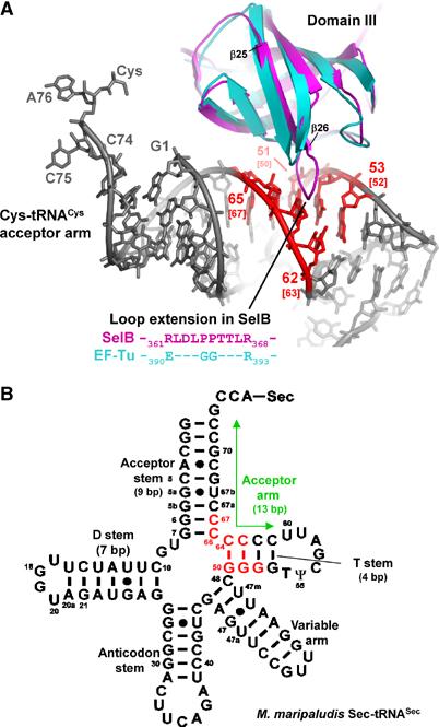
Figure 5.

Superposition of SelB domain III with the corresponding EF-Tu domain, which is involved in a tRNA backbone contact. (A) SelB (magenta) contains a loop that is considerably extended in comparison with EF-Tu (cyan), where this region is involved in tRNA (grey) binding. Contacts between SelB and the modelled tRNACys are coloured in red. The base-pair numbering is according to tRNACys. In addition, the corresponding bases from M. maripaludis tRNASec are shown in brackets. (B) Possible contact sites of SelB with tRNASec are shown in the secondary structure diagram of M. maripaludis Sec-tRNASec and are coloured in red. The contact area is derived from the tRNACys:SelB model. Note the prolonged 13 bp acceptor arm (labelled with a green arrow) that is formed by stacking of the 9 bp acceptor stem and the 4 bp T stem and is an important difference when compared to canonical elongator tRNAs.
