. 2017 May 9;6:e22904. doi: 10.7554/eLife.22904

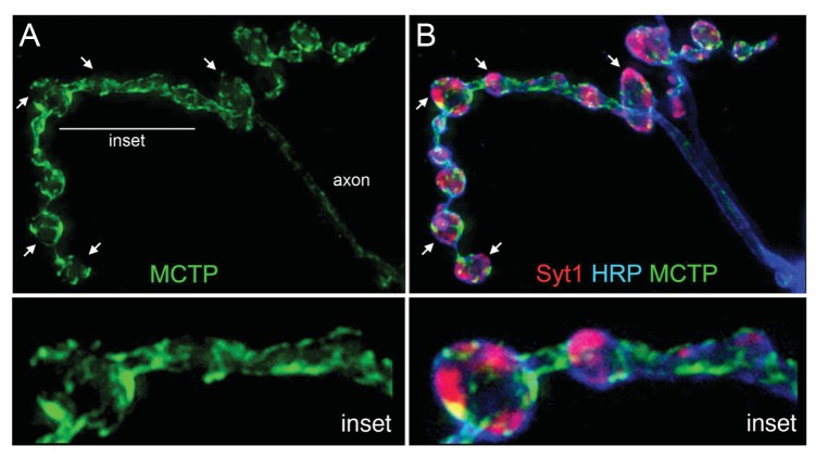
Figure 4. MCTP does not co-localize with the synaptic vesicle marker Syt1.

(A) Synaptic distribution of MCTP-Flag (green) at the NMJ. (B) The synapse in (A) co-stained with markers of the plasma membrane (anti-HRP, blue) and synaptic vesicles (anti-Syt1, red). Arrows indicate regions where Syt1 immunoreactivity is abundant and MCTP-Flag is absent. Insets (below; 2.5x magnification) of the region indicated by a horizontal line (top left) reveal regions occupied by Syt1 staining that are devoid of MCTP-Flag at higher magnification. Additional analysis of NMJ bouton anatomy and cytoskeleton is presented in Figure 4—figure supplement 1.

DOI: http://dx.doi.org/10.7554/eLife.22904.005

Figure 4.

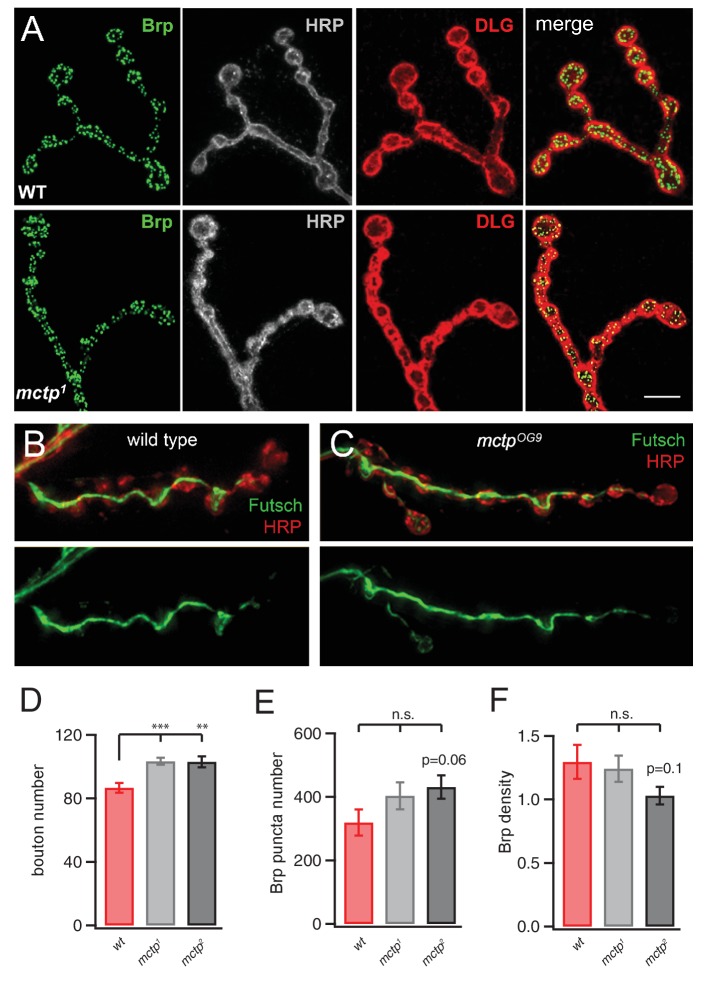
Figure 4—figure supplement 1. Normal NMJ Development in mctp mutants.

Figure 4—figure supplement 1.

(A) Representative images of the wild type (top) and mctp1 (bottom) NMJ stained with anti-Brp (green), HRP (white) and anti-Dlg (red). The far right images in each row represent the merged channels. Scale 5 µm. (B) Image of the NMJ stained with anti-Futsch (green) to label the synaptic microtubule cytoskeleton and HRP (red) to label the presynaptic membranes. Below, the anti-Futsch channel is shown in isolation. (C) Staining as in (B) for an NMJ in the mctpOG9 mutant. There is no qualitative difference in anti-Futsch staining. (D–F) Quantification of bouton number (D), anti-Brp puncta number per NMJ (E) and the density of Brp puncta per bouton (F). There is a small but statistically significant increase in bouton number in both the mctp1 and mctp2 mutants compared to wild type. There is no change in total Brp puncta number and a small decline in Brp density per bouton in mctp2 compared to wild type, consistent with the modest increase in bouton number without a change in active zone number, as defined by the presence of a Brp puncta.