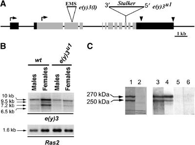
Figure 1.

The structure of e(y)3 gene and the nature of e(y)3 mutations. (A) Molecular structure of e(y)3 gene and transcripts. Gray boxes indicate the coding regions. Black boxes indicate 5′- and 3′-untranslated regions. Two alternative transcription start sites are shown by bent arrows; the alternative polyadenylation sites are shown by arrowheads. The Stalker insertion (position 4956 from the beginning of the longest ORF) in the e(y)3u1 allele and the site of 11-nt deletion at position 3525 in the e(y)3EMSl allele are indicated (not to scale). Both mutations lead to stop codon formation. The probe corresponding to the second exon was used for Northern hybridization (panel B). (B) Transcription of e(y)3 in wild-type and e(y)3u1 flies. The level of e(y)3 transcription is decreased in mutant males and females. Ras2 was used for normalization. The e(y)3 transcripts did not change in length in mutated flies, because splicing between the 3′ end of exon 9 and the sequences of Stalker 5′LTR resulted in replacement of 24 nt of exon 10 by 23 nt of Stalker. This produced a stop codon 85 amino acids downstream of the place of Stalker insertion. (C) Western blot detection of SAYP in embryonic nuclear extract. The lanes were developed with (1) nonpurified antiserum 1, (2) antiserum 1 after 1-h incubation with the peptide used for immunization, (3, 4) affinity-purified Ab1 and Ab2, and (5, 6) preimmune serum. Ab1 were raised against residues 102–308. Ab2 were raised against residues 495–643.
