Figure 1.

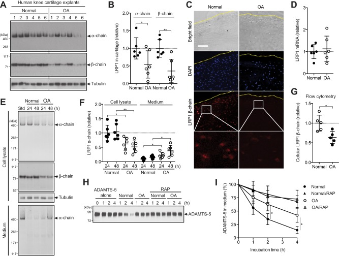
Increased ectodomain shedding of low‐density lipoprotein receptor–related protein 1 (LRP‐1) and reduced endocytic capacity of chondrocytes in human osteoarthritic (OA) cartilage. A, Western blotting of total proteins extracted from cartilage explants of human knee joints of OA patients and patients without arthritis (n = 6 each) with antibodies against α‐ and β‐chains of LRP‐1. B, Densitometric analysis of LRP‐1 in A. Data were normalized against tubulin. C, Immunofluorescence staining of LRP‐1 in frozen section of human knee cartilage. Dotted lines indicate the articular cartilage surface. Bar = 100 µm. D, Relative levels of mRNA for LRP‐1 in cartilage, measured by TaqMan quantitative reverse transcriptase–polymerase chain reaction. E, Representative Western blotting of LRP‐1 protein in cell lysates and medium of human normal and OA chondrocytes (n = 6 donors each). Std = standard cell lysates. F, Quantification of LRP‐1 α‐chain detected in E. G, Flow cytometric analysis of LRP‐1 β‐chain. H, Representative Western blotting for endocytosis of ADAMTS‐5 (10 nM) by human normal and OA chondrocytes (n = 3 donors each) with or without the LRP‐1 ligand antagonist receptor‐associated protein (RAP) (500 nM). ADAMTS‐5 in the medium was detected by Western blotting using anti–ADAMTS‐5 antibody. I, Quantification of findings in H. In B, D, F, and G, symbols represent individual cartilage donors; bars show the mean ± SD. The mean values in normal cartilage and chondrocytes were set at 1 (dashed lines). In I, values are the mean ± SD. ∗ = P < 0.05; ∗∗ = P < 0.01, by Student's 2‐tailed t‐test.
