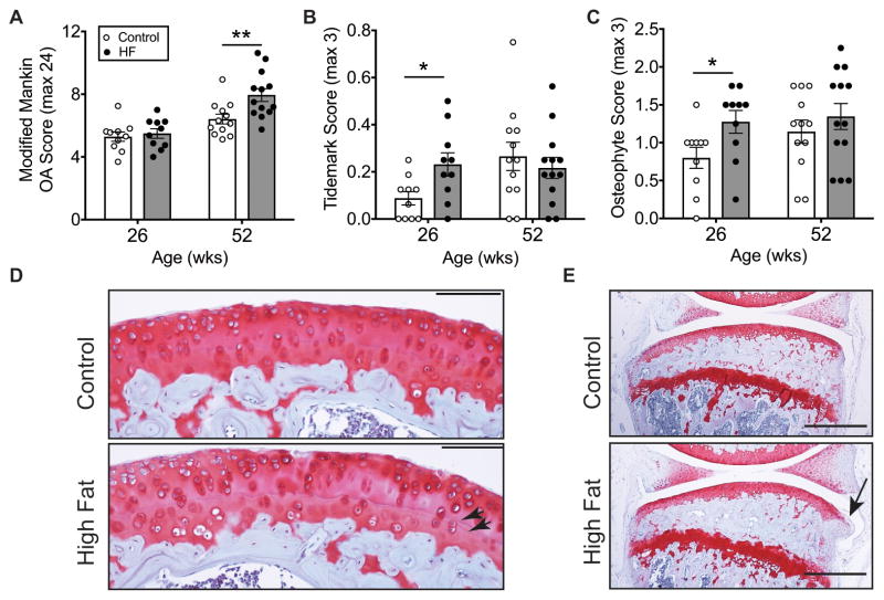
Figure 1. Age-dependent knee OA pathology in high-fat diet-induced obese mice.
Animals were fed either a 10% kcal fat control diet (Control) or a 60% kcal fat high-fat diet (HF) beginning at 6 weeks of age. (A) 46 weeks of a HF diet, but not 20 weeks, increased cartilage OA severity. OA was assessed by the knee cartilage modified Mankin OA score averaged for multiple sections and sites throughout the joint, including the medial and lateral tibia and femur. (B) 20 weeks of a HF diet increased duplication of the tidemark separating the uncalcified and calcified cartilage. After 46 weeks, however, both diet groups were similar. (C) 20 weeks of a HF diet also accelerated the development of tibial osteophytes. However, after 46 weeks, Control and HF diet groups were no longer different. (D) 200X magnification sagittal image of the medial tibia. Arrowheads indicate tidemarks; scale bar = 100μm. (E) 40X magnification sagittal image of the medial compartment of the knee in mice fed a control or HF diet for 20 weeks. Arrow indicates an osteophyte; scale bar = 500μm. Values are mean ± SEM for n=10 per diet (26 wks) and n=12 Control and n=13 HF (52 wks); *p<0.05, **p<0.01.

