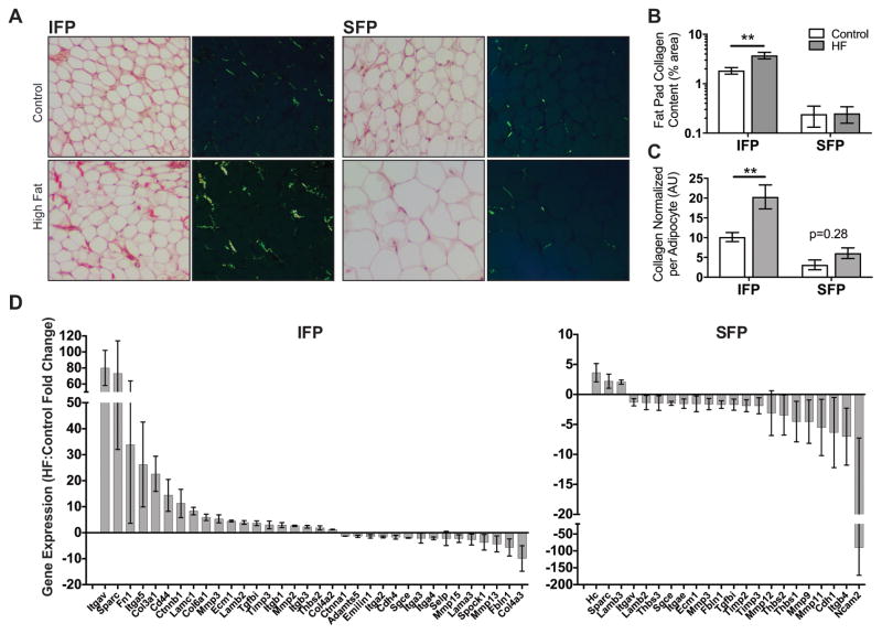
Figure 5. HF diet-induced infrapatellar and subcutaneous adipose tissue fibrosis and extracellular matrix gene expression.
(A) Brightfield and epipolarized UV light images of Sirius Red-stained IFP and SFP paraffin sections from mice fed a control or HF diet for 20 weeks. Yellow-green pixels from epipolarized images were quantified to determine adipose tissue collagen content. (B) HF diet significantly increased IFP collagen content but did not alter SFP collagen content. (C) Fat pad collagen content was normalized to the average adipocyte area per fat pad to adjust for anatomical and HF diet-associated differences in adipocyte size. Even with normalization, IFP but not SFP collagen content was elevated by a HF diet. n = 5 per diet. (D) Gene expression was measured by RT2 Profiler™ PCR Extracellular Matrix and Adhesion Molecules targeted array following 20 weeks of a HF diet. Significant differentially expressed genes due to a HF diet are plotted from highest to lowest for the IFP and SFP (p<0.05 based on 95% confidence interval (CI) of fold-change value). Values are mean ± 95% CI; values are also reported in Table S2. Sample sizes same as in Figure 4. **p<0.01.

