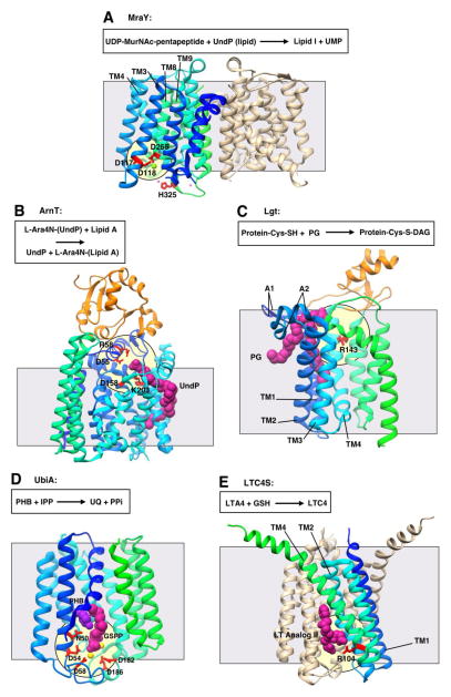
Figure 3. Representative structures showing Interfacial catalysis (II).
All structures are shown in ribbon representation with bound ligands shown in CPK representation. Transmembrane domains are shown in gradient coloring from the N-terminus to the C-terminus (blue to cyan to green), while extramembrane soluble domains are shown in orange. The grey square shows the approximate location of the membrane border. The pale yellow circle indicates the location of the putative active site. All structures oriented with the cytosolic face of the membrane on the bottom. A. Phospho-MurNAc-pentapeptide translocase (MraY; PDB 4J72) with putative catalytic residues in red. Nickel ion in pink, magnesium ion in light green. UDP, uridine diphosphate; UMP uridine monophosphate; UndP, undecaprenyl phosphate. B. A4-amino-4-deoxy-L-arabinose transferase (ArnT; PDB 5F15) with putative catalytic residues in red. UndP in magenta. C. Lipoprotein diacylglycerol transferase (Lgt; PDB 5A2C) with putative catalytic residues in red. Phosphatidylglycerol (PG) in magneta. D. Prenyltransferase UbiA (PDB 4OD5) with putative catalytic residues in red. Geranyl thiolopyrophosphate (GSPP) in magenta, p-hydroxybenzoate (PHB) in purple, magnesium ions in yellow. A1 and A2, arm 1 and arm 2; IPP, isoprenylpyrophosphate; UQ, ubiquinone. E. Leukotriene C4 Synthase (LTC4S; PDB 4J7T) with putative catalytic residue in red. Leukotriene (LT) analog in magenta. LTA4, (5S)-trans-5,6-oxido-7,9-trans-11,14-cis-eicosatetraenoic acid; LTC4, (6R)-S-glutathionyl-7,9-trans-11,14-cis-eicosatetraenoic acid ; GSH, glutathione.

