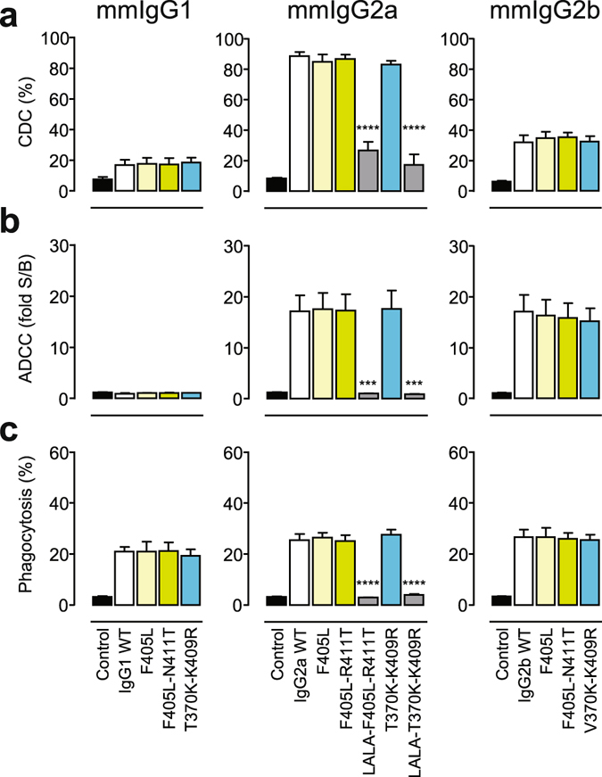
Figure 4.

(a) CDC of Raji cells incubated with a fixed 10 μg/mL concentration of the indicated mmIgG1 (left), mmIgG2a (middle), or mmIgG2b (right) variants of mAb 7D8 in the presence of 20% (v/v) pooled human serum. (b) Surrogate ADCC activity of Jurkat-NFAT-mFcγRIV effector (E) cells induced by Raji target (T) cells incubated with a fixed 3 μg/mL concentration of the indicated variants (see A), with an E:T ratio of 1:1. (c) Phagocytosis of Daudi cells (T) by bone marrow-derived mouse macrophages (E) incubated with a fixed 1 μg/mL concentration of the indicated variants (see A), with an E:T ratio of 1:1. Data represent mean ± SEM of at least three experiments. Statistical significance (compared to WT) was determined by one-way ANOVA (***P < 0.001; ****P < 0.0001).
