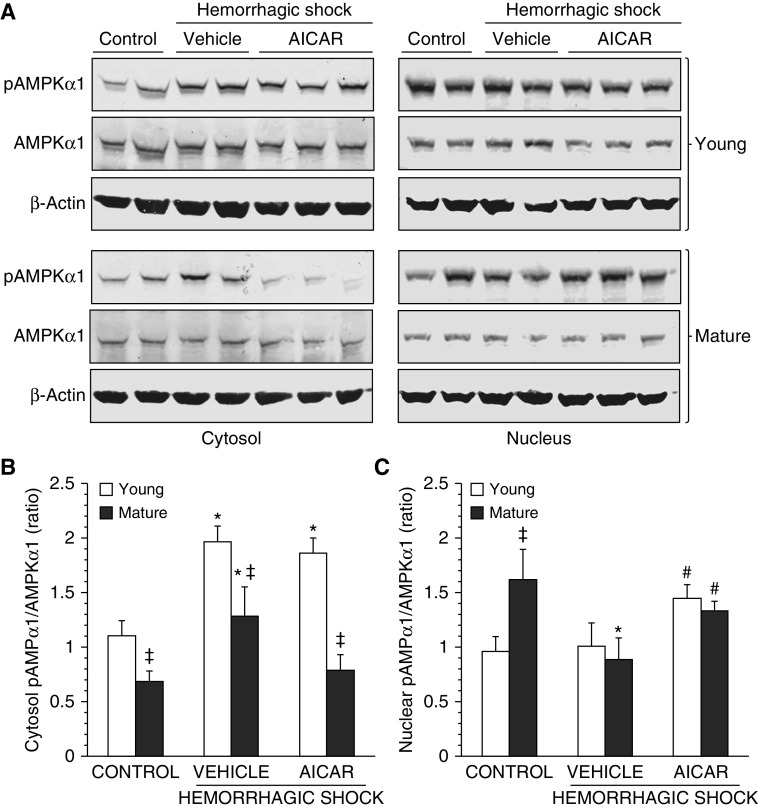
Figure 7.
(A) Western blot analysis of active phosphorylated AMPK (pAMPK) α1, AMPKα1, and β-actin (used as loading control protein) in lung cytosol and nuclear extracts. Image analyses of pAMPKα1/AMPKα1 ratio as determined by densitometry (B) in the cytosol and (C) in the nucleus. Data are expressed as mean ± SEM of four to six animals for each group and are expressed as ratio of relative intensity units. *P < 0.05 versus age-matched control mice; #P < 0.05 versus vehicle-treated group of the same age; ‡P < 0.05 versus young group.

