Figure 5.
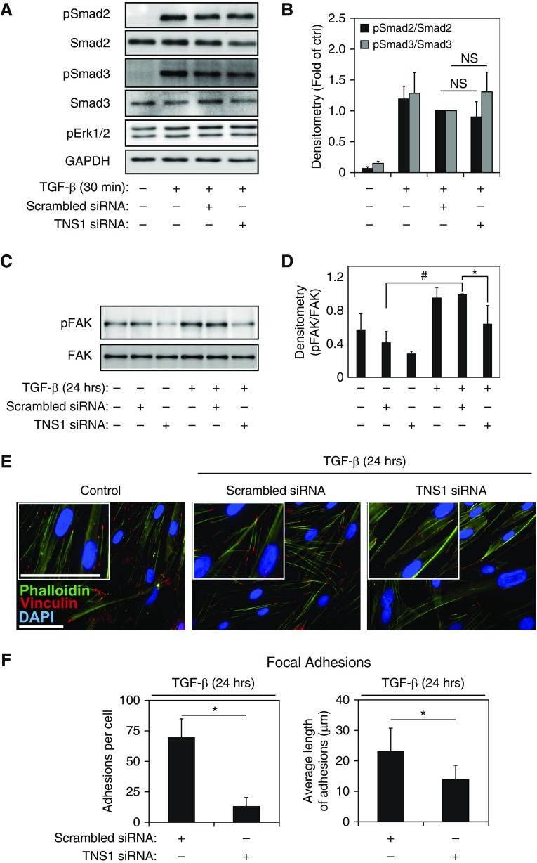
TNS1 is dispensable for TGF-β signaling but required for focal adhesion–dependent signals. (A and B) HLFs were transfected with TNS1 siRNA or scrambled control followed by treatment with 1 ng/ml TGF-β for 30 minutes and Western blotting and densitometry for the indicated (phospho) proteins. Mixed-effect ANOVA (*P < 0.05; NS, not significant) was used for statistical analysis. (C and D) HLFs were transfected with TNS1 siRNA or scrambled control, followed by treatment with 1 ng/ml TGF-β for 24 hours, Western blotting, and densitometry for the phosphorylated tyrosine 397 residue of focal adhesion kinase (FAK). Mixed-effect ANOVA (* and #P < 0.05) was used for statistical analysis. (E) Merged immunocytochemistry images of HLFs stained against phalloidin (green) and vinculin (red) upon TNS1 knockdown using TNS1 siRNA (or treatment with scrambled control) and treatment with 1 ng/ml TGF-β for 24 hours. Scale bar, 50 μm. Insets show digital magnifications of vinculin staining for each condition. Scale bar, 50 μM. (F) Quantitation of number and length of focal (vinculin-containing) adhesions in TGF-β–induced (1 ng/ml for 24 h) myofibroblasts under siRNA-mediated TNS1 knockdown or scrambled control. Student’s t test (*P < 0.05) was used for statistical analysis. * and # symbols designate significant difference between the indicated conditions. Data are presented as means (±SD). DAPI, 4′,6-diamidino-2-phenylindole.

